Ovule
Structure of Ovule:
Ovule is an integumented megasporangium found in sper- matophytes which develops into seed after fertilisation. An angiosperm ovule is typically an ovoid and whitish structure. It occurs inside ovary where it is attached to a parenchymatous cushion called placenta either singly or in a cluster.
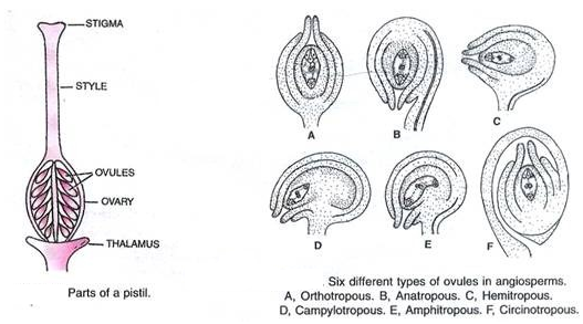
The ovule is stalked. The stalk is called funiculus or funicle. The point of attachment of the body of the ovule with the funiculus is known as hilum. Depending upon the configuration and orientation of the body of ovule in relation to funiculus, there are six types of ovules— orthotropous (atropous, erect), anatropous (inverted), hemitropous (half inverted), campylotropous (body curved), amphitropous (both body and embryo sac curved) and circinotropous (funiculus coiled around the ovule).
In the typical (anatropous) ovule the funiculus is fused with body of the ovule lengthwise beyond the hilum. It gives rise to a longitudinal ridge called raphe. Funiculus contains a vascular strand for the supply of nourishment to the ovule.
The body of the ovule consists of a mass of parenchymatous cells named nucellus. It is equivalent to mega sporangium. Nucellus may be quite massive (crassinucellate ovule) or thin (tenuinucellate ovule). It is surrounded by one (unitegmic ovule, e.g., higher dicots) or two (bitegmic ovule, e.g., monocots and primitive dicots) multicellular integuments.
Rarely an ovule may be surrounded by three integuments (tritegmic, e.g., Asphodelus) or the integuments are absent (ategmic, e.g., Santalum). Free surfaces of nucellus and integuments are covered by cuticle. The integuments leave a narrow pore or passage at one end of the ovule. It is known as micropyle. The place of origin of the integuments usually lies at the opposite end. It is termed as chalaza.
Female gametophyte or embryo sac is embedded in the micropylar half of the nucelus.
Development of Ovule (Me- gasporogenesis):
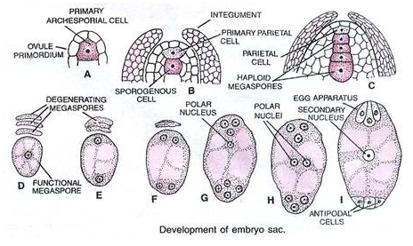
Ovule develops as primordium and then mound of nucellus over placenta. Initials of integuments develop from its base. They grow and come to surround the nucellus on all sides except at the tip or micropylar region. In the hypoder- mal region of nucellus towards the micropylar end develops a primary archesporial cell. It grows in size and develops a prominent nucleus.
The archesporial cell often divides once into outer primary parietal or wall cell and inner primary sporogenous cell. Primary parietal cell may divide one or more times. The primary sporogenous cell commonly functions directly as diploid megaspore mother cell or megasporocyte.
The megaspore mother cell (MMC) undergoes meiosis and forms a linear tetrad of 4 haploid megaspores. The process of meiotic formation of haploid me- gaspores from diploid megaspore mother cell is called megasporogenesis. Commonly the chalazal megaspore remains functional while the other 3 degenerate.
Development of Female Gametophyte (Mega-gametogenesis):
The functional megaspore is the first cell of female gametophyte. The cell enlarges and undergoes three free nuclear mitotic divisions. The first division produces two nucleate embryo sac. The two nuclei shift to the two ends and divide there twice forming four nucleate and then eight nucleate structure.
One nucleus from each side moves to the middle. They are called polar nuclei. The remaining three nuclei form cells at the two ends, 3 celled egg apparatus at the micropylar end and three antipodal cells at the chalazal end.
The middle bi-nucleate part organises itself into central cell. Embryo sac developed from a single megaspore is called monosporic.